Endomembrane System
Endoplasmic Reticulum - It is discovered by Porter et al and Thompson in 1945. The name was given by Porter in 1953.It is a system of membrane lines channels found in all eukaryotic cells except mature erythrocytes. It constitutes of more than 30 to 60% of the cells. It is known as different name in different organelles. This are sarcoplasmic reticulum in muscle cells, myeloid bodies in at the base of retinal pigments cell, nissl granules which are believed to be formed from E.R.
Structure of Endoplasmic Reticulum - It is a membrane bound organelles whose membranes are thinner than other cell membrane. It is often connected with the outer membrane of nuclear envelope and open at plasmalemma. It is made up of tubules in adipose tissue, few vessicles in spermatocytes, reticulocytes and is best developed in metabolically active cells called liver, pancreas ,plasma cells, intestinal cells, fibroblasts. Endoplasmic reticulum is made up of three parts –cisternae, tubules and vessicles. Membranes of endoplasmic reticulum contains ribosome and is called Rough endoplasmic reticulum (RER). This attachment between ribosome- ER is done by glycoproteins (ribophorin 1 and ribophorin 2). RER often contains minute pores below ribosome to pass and synthesised polypeptides into the lumen for transport ER without attached ribosomes is called agranular or smooth endoplasmic reticulum (SER). It is associated with synthesis of vitamin, carbohydrate, fat, sterol and detoxification.RER is abundant in cisternae, and in the cells that are engaged in production and excretion of proteins (plasma cells, goblet cells, pancreatic acinus cell and in certain liver cells). Broken pieces of endoplasmic reticulum are appeared as microsomes.

Enzymes found in Endoplasmic Reticulum - Enzymes are found in two different areas of the endoplasmic reticulum. They are occur in – cytoplasmic surface (P-450, cytochrome b5, some reductase and nucleotidase), luminaries surface (glucose 6 phosphatase, peptidase, beta glucoronidase).
Golgi Apparatus - This organelles is a complex organisms which is made up of membrane like stack of cisternae , network of tubules , vessicles and vacuoles that were first discovered by George in 1867 and studied by Camillio Golgi in 1898 in cells of Barn Owl and Cat through metallic impregnation techniques.
Structure of Golgi Apparatus - Golgi apparatus is found in ever eucaryotic cell except RBC and sieve tubes. It is also absent in prokaryotes and sperm cells of seedless embryophytes. In plant cell it is made up of different isolated units called dictyosome but in animal cells it occurs in single compact or loose complex. A unit of Golgi complex is called golgisome. Golgi apparatus are surrounded by a clear zone of exclusion in which mitochondria, ribosome, plastids , storage granules are absent. A golgisome or dictyosome has a central stack which are 3to 10 curved parallel membrane bound narrow called cisternae. Cisternae are interconnected with each other. Golgi apparatus has two faces one is distal maturing and proximal forming face (convex or CIS face). Cis face receives materials from endoplasmic reticulum and cytosol while concave face maturing face gives out large Golgisome vacuoles and small vessicles having transformed materials. Vessicles are developed from tubules.Membranes of proximal cisternae are thin (50 to 60 angstroms).They progressively become thick towards the distal side ,reaching a thickness of 75 to 80 anstrom. It receives materials from cytosol and endoplasmic reticulum in the form of transitional vessicles.
It produces materials for secretion, takes part in transformation of membrane, formation of glycoproteins, complex heteropoly saccharide, hormone, Melanine, matrix of connective tissue.

Lysosome - It is also called suicide bags or disposal
units. It contains hydrolysis enzymes and surrounded by single membrane.This
are small vessicles whose size is 0.2 to 0.8 micrometres.It was discovered by
Christian de Duve in 1955 but we’re named and observed under microscope by Novikoff
in 1956.They are most abundant in phagocytic cell (WBC), chondroclasts and
osteoclasts. This are formed by Golgi apparatus and contain some (40 types ) acid
hydrolase for digestion of various materials (nucleases, proteases, lipases,
glucosidase, phosphatases, sulphatases). Membrane of lysosome is strengthen by
cortisone, cortisol, anti histamine, heparin, chloroquine and a type of
cholesterol.They are known as membrane stabilizer. It becomes fragile in absence of oxygen or
presence of vitamin A, vitamin E, progesterone, testosterone,bile salts, high
energy radiations. This are called membrane labilizers. Lysosomes are useful in
metamorphosis (eating away larval organs), removal of obstructions,
intracellular scavenging, osteogenesis, hormone elaboration, fertilization etc.
Lysosomes causes inflammation and collagen synthesis. Lung fibrosis is produced
in same way. In many plants lysosome functions are performed by spherasomes and
vacuoles.

Polymorphism of Lysosome - Lysosome show polymorphism. Primary Lysosome are newly formed protolysosomes that are storage granules, heterolysosomes are digestive vacuoles which consists of primary Lysosome and phagosome, residual lysosome are having undigested material which undergo ephagy, autophagic vacuoles causes autolysis of useless part. Secondary lysosome is consists of digestive vacuoles, autophagic vacuoles, residual vacuoles.
Vacuoles - These are noncytoplaamic sacs that are separated from the cytoplasm by membrane. Vacuolea are mainly found in plant cell. But in animal cells it is also observed very few in number. Plant vacuoles are bigger in size than animal cell vacuoles. Vacuoles are of four types-
1. Sap Vacuoles – These are enclosed sap or water with dissolved inorganic and organic substances. A sap vacuole is surrounded by a membrane which is known as tonoplast. Plants contain this in large size and animals cell in small size and numerous in number.
These vacuoles maintain osmotic pressure and turgidity .It is filled of usefully and waste materials.
2. Contractile Vacuoles - This occur in some simple fresh water forms organisms like Amoeba, Paramecium, Chlamydomonas.These vacuoles are surrounded by a few feeding canals . It regulates osmoregulation and excretion.
3. Food Vacuoles - It is a complex of lysosome and phagosome and digestion occurs inside them.
4. Gas Vacuoles – It is also called pseudovacuoles. Gas or air vacuoles occur in some prokaryotes. Each of them is made up of large number of gas vessicles which are hexagonal and submicroscopic. A gas vessicles is surrounded by a thin protein membrane . It stores metabolic gases and take part in buoyancy regulations.
You might like these
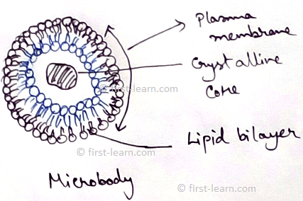
Explain Microbodies | Definition | Glyoxisomes|Glycosome|Hydrogenosome
This in 1954 first was the first to observed microbodies. Microbodies are spherical or oval in shape with the diameter of 0.2 to 1.5 micrometer. It has outer covering with lipid and lipoprotein membrane. It contains different enzymes for catalase but do not contain any
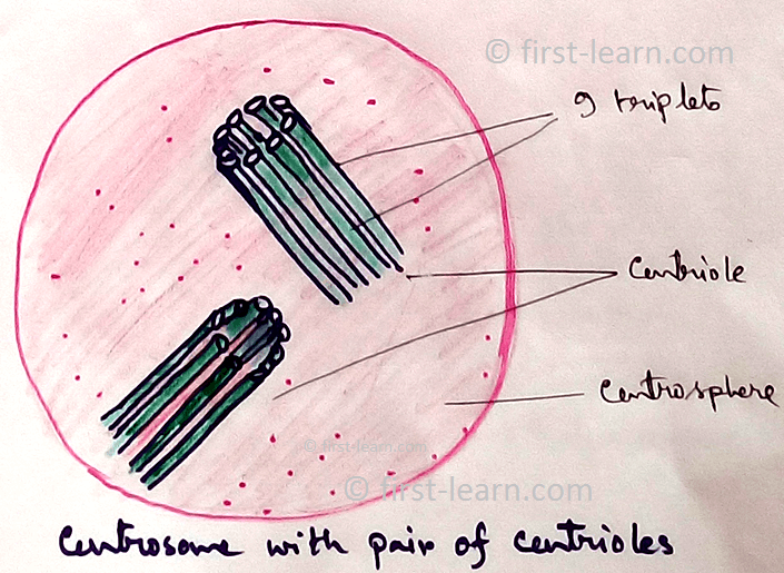
Centrosome and Centrioles | Distribution of the Centrosome | Structure
Definition of Centrosome - Centrosomeis a more or less spherical mass of dense cytoplasm situated close to the nucleus and contains a pair of cylindrical bodies which are concerned with spindle formation during cell division. In 1888 Boveri first discovered these organelles
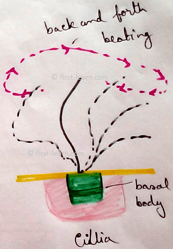
Cilia and Flagella | Definition | Location | Origin |Types of Flagella
Definition – Cilia and flagella are hair like micro tubular organelles projecting from the cell surface into the extra cellular medium and are concerned with cell motility.
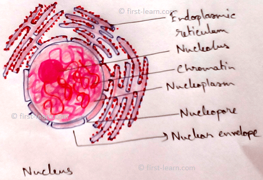
Explain about Nucleus | Nuclear Envelope | Nucleoplasm |Nuclear matrix
Robert Brown in 1831 first discovered a double membrane covered protoplasmic body that contains hereditary information. A cell usually contains a single nucleus (uninucleate, monokaryote).
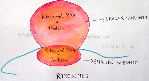
Explain about Ribosomes | Distribution and Origin of Ribosomes
Ribosomes are small ,granular, non- membranous structure which is mafe up of RNA and protein and concerned with protein synthesis. In 1943 presence of ribosomes are first observed by Claude and later in 1955 Palade described a detailed description of it by define the name.
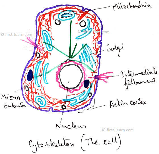
Cytoskeletal Structure | Functions and Types of Cytoskeleton Structure
Cytoskeletal structures are fibrous or fine tubular structures that consists of supportive structure of cell. The term cytoskeletal is coined by Koltzoff in 1928.
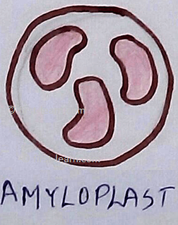
Define the Plastids | Structure of Plastids | Types of Plastids
Plastids are semi-autonomous cell organelles which are surrounded by double membrane envelope, take part in storage and synthesis of organic compounds which occurred in some plant and protistans.Plastidome consists of all the whole plastids complex of cell.
Explain about Mitochondria | Functions of Mitochondria | Distribution
The term mitochondria can be broken into mito and chondrion. The term “mito “ means fibril and” chondrion “means granule. Definition of mitochondria - The rod like filamentous, spherical or oval, double membrane bound cytoplasmic organelles of eukaryotic cells ,which are
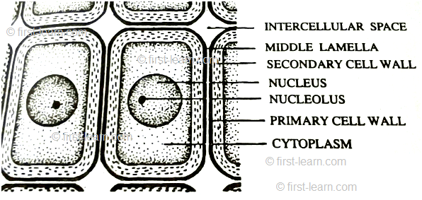
Cell Wall | Definition of Cell Wall |Middle Lamella|Primary, Secondary
Definition of cell wall- The thick and rigid , nonliving covering present just outside the plasma membrane of plant cells is called cell wall. It is discovered by Robert Hooke in 1665 while observing cell in the section of cork.
Explain about Cell Membrane | Structure of the Cell Membrane
Definition - Cell membrane is the thin, elastic and semipermeable living membrane that surrounds the protoplasm of a cell is called cell membrane o plasma membrane or plasma lemma.

From Endomembrane System to HOME PAGE


Recent Articles
-
Plants Around Us | Big & Small Plants | Shrubs & Herbs | Water Plants
Feb 03, 26 02:01 AM
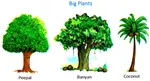 We see different types of plants around us. Plants are living things. They breathe and grow. They also reproduce. Most of the plants grow on land. Some plants grow in water.
 -
Formed Elements of Blood | Erythrocytes | ESR |Leukocytes |Neutrophils
Jan 15, 26 01:25 AM
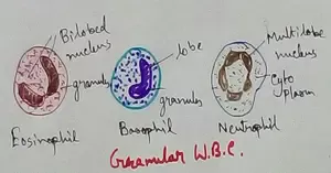 Formed elements formed elements are constitute about 45 % of blood afeias haematocrit value packed cell volume mostly of red blood corpuscles and are of 3 types- erythrocytes, leukocytes and blood pla…
 -
What Is Plasma? | Blood Plasma | Proteins | Nutrients | Cholesterol
Nov 07, 25 10:29 AM
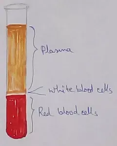 Blood is a mobile fluid which is a connective tissue and is derived from the mesoderm like cell any other connective tissue. Colour of blood is reddish and that flows inside the blood vessels by means…
 -
Disorders of Respiratory System | Tuberculosis | Pleurisy | Emphysema
Oct 28, 25 11:39 PM
Tuberculosis is very common disease and is caused by a type of bacteria called Mycobacterium tuberculosis. This disease causes different trouble in the respiration and infection of several parts of th… -
Regulation of Respiration | Respiratory Centres | Inspiratory Area |
Oct 14, 25 12:13 AM
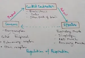 Respiratory Centre is the area that controls the rate of respiration and it is observed to be located in medulla oblongata and pons. Respiratory Centre has the following will dispersed components like…
New! Comments
Have your say about what you just read! Leave me a comment in the box below.