Human Cheek Cells Under the Microscope
Staining of human cheek cell - First we have to take a clean piece of cotton swab and scrap the epithelium layer from the inside of our mouth. It is then put on the previously cleaned slide and smear is prepared. Then a staining solution called haematoxylin is added as a colour solution. Then the excess stain is removed and cover slip that is previously cleaned is added to the slide to make it appropriate for looking under microscope. Then the cheek cells are observed under microscope.
Human cheek cells are observed under microscope-
1. Cells are polygonal or flat in shape and structure –
2. They have irregular cellular thin boundaries which contains jelly like cytoplasm and the cytoplasm are granular.
3. This cell do not have plastids, vacuoles or cell wall.
4. They are generally made up of squamous epithelium cells.

Cell Wall – As animal cells do not contain any cell wall .So the cheek cell of the human embryo do not contain any cell wall.
Cell Membrane – Cell membrane are very thin border of the cells of the animal.
Question and Answer on Human Cheek Cells Under the Microscope
1. What is the name of the stain that is used for staining human cheek cell?
Answer: Haematoxylin is used for staining.
You might like these
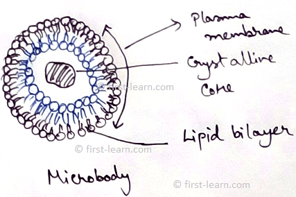
Explain Microbodies | Definition | Glyoxisomes|Glycosome|Hydrogenosome
This in 1954 first was the first to observed microbodies. Microbodies are spherical or oval in shape with the diameter of 0.2 to 1.5 micrometer. It has outer covering with lipid and lipoprotein membrane. It contains different enzymes for catalase but do not contain any
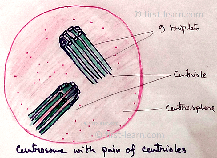
Centrosome and Centrioles | Distribution of the Centrosome | Structure
Definition of Centrosome - Centrosomeis a more or less spherical mass of dense cytoplasm situated close to the nucleus and contains a pair of cylindrical bodies which are concerned with spindle formation during cell division. In 1888 Boveri first discovered these organelles
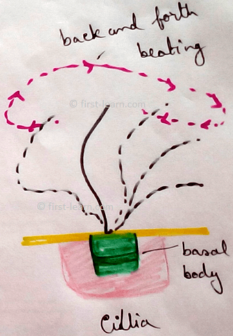
Cilia and Flagella | Definition | Location | Origin |Types of Flagella
Definition – Cilia and flagella are hair like micro tubular organelles projecting from the cell surface into the extra cellular medium and are concerned with cell motility.
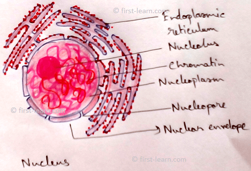
Explain about Nucleus | Nuclear Envelope | Nucleoplasm |Nuclear matrix
Robert Brown in 1831 first discovered a double membrane covered protoplasmic body that contains hereditary information. A cell usually contains a single nucleus (uninucleate, monokaryote).
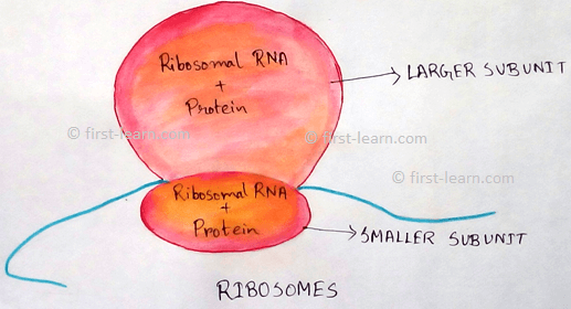
Explain about Ribosomes | Distribution and Origin of Ribosomes
Ribosomes are small ,granular, non- membranous structure which is mafe up of RNA and protein and concerned with protein synthesis. In 1943 presence of ribosomes are first observed by Claude and later in 1955 Palade described a detailed description of it by define the name.
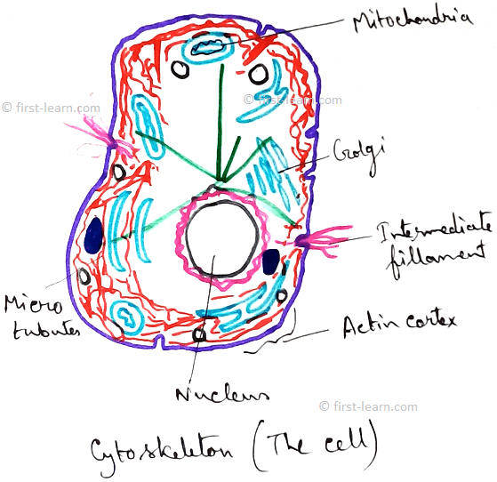
Cytoskeletal Structure | Functions and Types of Cytoskeleton Structure
Cytoskeletal structures are fibrous or fine tubular structures that consists of supportive structure of cell. The term cytoskeletal is coined by Koltzoff in 1928.
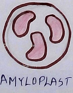
Define the Plastids | Structure of Plastids | Types of Plastids
Plastids are semi-autonomous cell organelles which are surrounded by double membrane envelope, take part in storage and synthesis of organic compounds which occurred in some plant and protistans.Plastidome consists of all the whole plastids complex of cell.
Explain about Mitochondria | Functions of Mitochondria | Distribution
The term mitochondria can be broken into mito and chondrion. The term “mito “ means fibril and” chondrion “means granule. Definition of mitochondria - The rod like filamentous, spherical or oval, double membrane bound cytoplasmic organelles of eukaryotic cells ,which are
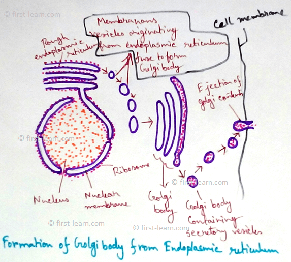
Endomembrane System | Endoplasmic Reticulum | Golgi Apparatus
Endoplasmic reticulum-It is discovered by Porter et al and Thompson in 1945. The name was given by Porter in 1953.It is a system of membrane lines channels found in all eukaryotic cells except mature erythrocytes.
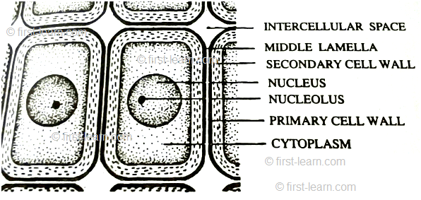
Cell Wall | Definition of Cell Wall |Middle Lamella|Primary, Secondary
Definition of cell wall- The thick and rigid , nonliving covering present just outside the plasma membrane of plant cells is called cell wall. It is discovered by Robert Hooke in 1665 while observing cell in the section of cork.

From Human Cheek Cells Under the Microscope to HOME PAGE


Recent Articles
-
Plants Around Us | Big & Small Plants | Shrubs & Herbs | Water Plants
Feb 03, 26 02:01 AM
 We see different types of plants around us. Plants are living things. They breathe and grow. They also reproduce. Most of the plants grow on land. Some plants grow in water.
 -
Formed Elements of Blood | Erythrocytes | ESR |Leukocytes |Neutrophils
Jan 15, 26 01:25 AM
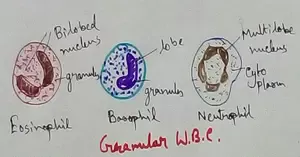 Formed elements formed elements are constitute about 45 % of blood afeias haematocrit value packed cell volume mostly of red blood corpuscles and are of 3 types- erythrocytes, leukocytes and blood pla…
 -
What Is Plasma? | Blood Plasma | Proteins | Nutrients | Cholesterol
Nov 07, 25 10:29 AM
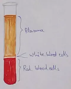 Blood is a mobile fluid which is a connective tissue and is derived from the mesoderm like cell any other connective tissue. Colour of blood is reddish and that flows inside the blood vessels by means…
 -
Disorders of Respiratory System | Tuberculosis | Pleurisy | Emphysema
Oct 28, 25 11:39 PM
Tuberculosis is very common disease and is caused by a type of bacteria called Mycobacterium tuberculosis. This disease causes different trouble in the respiration and infection of several parts of th… -
Regulation of Respiration | Respiratory Centres | Inspiratory Area |
Oct 14, 25 12:13 AM
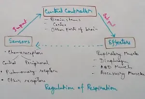 Respiratory Centre is the area that controls the rate of respiration and it is observed to be located in medulla oblongata and pons. Respiratory Centre has the following will dispersed components like…
New! Comments
Have your say about what you just read! Leave me a comment in the box below.