Onion Peels Observed Under the Microscope
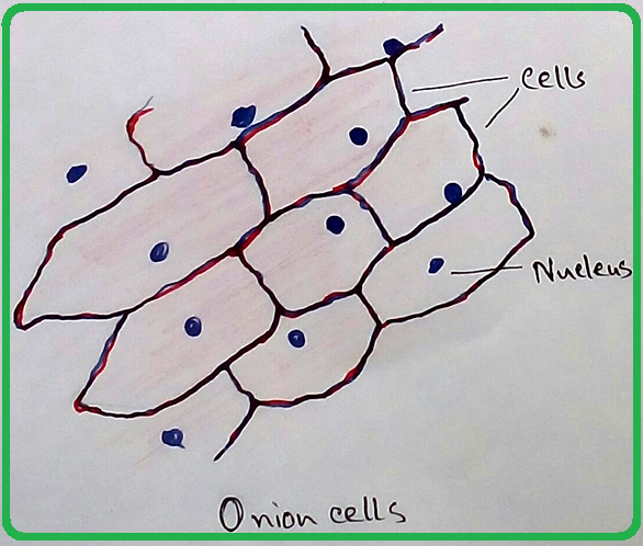
Cells present in onion peel can be observed
under microscope. For this onion peels are first isolated. For this experiment outer
most scale of the onion is removed and is cut into four equal halves. It is a
monocot plant. Then with the help of a pairs of forcep the scale of onion is
peeled out. It should be fleshy unless in the dried ones experiment cannot be
done due to the absence of proper cellular structure. Then the onion peel is
sized into 5mm × 5mm size (approx). A slide should be properly cleaned and keep
it for the preparation of the onion peel.
Then the part of the onion peel is placed on the slide and a previously cleaned cover slip is placed on the part of the peel. During the placement of cover slip precautions should be taken that there should not be present of air bubbles. It is stained with iodine solutions or eosin solutions. Eosins are responsible for staining of nucleus as it is acidic in nature. After adding of stain by the help of dropper excess stain should be removed by washing. Then the prepared slide can be observed under microscope.
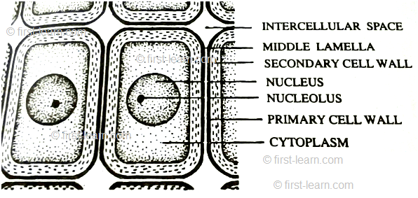
Observation under Microscope - Cells are appeared to be prominent, individual, linear, rectangular in shape. If it is observed under electron microscope in low resolution then the presence of cell wall and nucleus are observed. But if it is observed under microscope in high resolution then presence of cell vacuoles can be observed properly.
Characteristics features of the onion peels are -
1. Cells are firmly bound to each other.
2. Nucleus present in the cells are slightly towards the periphery of the cells. Which is the one of the confirmation point of onion peel.
From Onion Peels Observed Under the Microscope to HOME PAGE
Recent Articles
-
Plants Around Us | Big & Small Plants | Shrubs & Herbs | Water Plants
Feb 03, 26 02:01 AM
We see different types of plants around us. Plants are living things. They breathe and grow. They also reproduce. Most of the plants grow on land. Some plants grow in water. -
Formed Elements of Blood | Erythrocytes | ESR |Leukocytes |Neutrophils
Jan 15, 26 01:25 AM
Formed elements formed elements are constitute about 45 % of blood afeias haematocrit value packed cell volume mostly of red blood corpuscles and are of 3 types- erythrocytes, leukocytes and blood pla… -
What Is Plasma? | Blood Plasma | Proteins | Nutrients | Cholesterol
Nov 07, 25 10:29 AM
Blood is a mobile fluid which is a connective tissue and is derived from the mesoderm like cell any other connective tissue. Colour of blood is reddish and that flows inside the blood vessels by means… -
Disorders of Respiratory System | Tuberculosis | Pleurisy | Emphysema
Oct 28, 25 11:39 PM
Tuberculosis is very common disease and is caused by a type of bacteria called Mycobacterium tuberculosis. This disease causes different trouble in the respiration and infection of several parts of th… -
Regulation of Respiration | Respiratory Centres | Inspiratory Area |
Oct 14, 25 12:13 AM
Respiratory Centre is the area that controls the rate of respiration and it is observed to be located in medulla oblongata and pons. Respiratory Centre has the following will dispersed components like…




New! Comments
Have your say about what you just read! Leave me a comment in the box below.